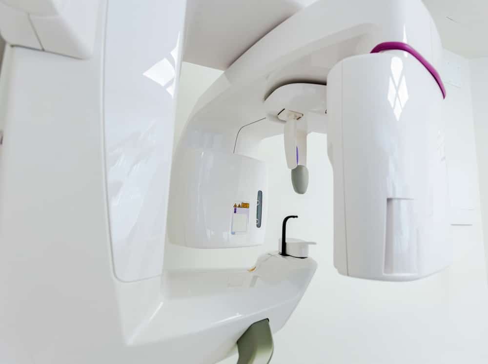
CBCT Imaging
Advanced dental care starts with accurate diagnostics. At Dental Spa of Orange in Orange, CA, our team uses cone beam computed tomography (CBCT) to capture high-resolution 3D images of your teeth, jaw, and surrounding structures. This imaging technology provides a level of detail beyond traditional dental x-rays, helping us evaluate concerns more clearly and plan treatment with greater confidence. Whether you are exploring dental implants, evaluating an impacted tooth, or troubleshooting ongoing symptoms, CBCT imaging helps us see the full picture, so care can be more precise and personalized.
What Is Cone Beam 3D Imaging?
CBCT is an advanced type of dental imaging that captures hundreds of images from multiple angles and assembles them into a detailed 3D view. Instead of providing a flat, two-dimensional image, CBCT creates a layered model that allows us to evaluate teeth, roots, bone, nerve pathways, and surrounding anatomy with greater clarity.
- Better visualization of jaw structure and bone density.
- Accurate mapping of anatomical features, such as nerve positioning.
- More confident planning for complex procedures.
- Comprehensive visuals: Produces a layered, 3D model of your oral anatomy for better evaluation.
- Fast and efficient: Captures a complete scan in seconds, right in the dental office.
- Comfortable experience: No special preparation, just a quick, non-invasive scan.
Because the scan captures detailed information in a single session, it can also reduce the need for multiple imaging appointments when more detail is required for treatment planning.
How CBCT Enhances Dental Diagnosis & Treatment
CBCT imaging helps reduce guesswork by providing detailed information about anatomy and tooth structures. This is especially important when planning procedures where precision matters, such as implant placement, surgical extractions, or complex root canal cases.
- Identify hidden concerns that may not show clearly on traditional imaging.
- Evaluate bone volume and density for tooth replacement planning.
- Map root structure and anatomy for more targeted care.
- Plan surgical procedures with improved visualization.
- Pinpoint accuracy: Helps guide precise procedures by mapping bone density and nearby anatomical structures.
- Fewer surprises: Detects hidden concerns such as impacted teeth, cysts, or infections before complications occur.
- Smarter decisions: Provides detailed insights that help tailor care to your individual needs.
- Bone support: Whether there is enough bone and density to support implants or other treatment goals.
- Anatomy location: Where important structures are positioned, so planning is safer and more predictable.
- Infection signs: Whether there are changes around root tips or in the surrounding bone that suggest inflammation.
- Tooth positioning: Whether impacted or shifted teeth require coordinated treatment planning.
- Complex structures: Whether the tooth anatomy calls for a more detailed approach to treatment.

Common Uses Of CBCT In Dentistry
CBCT supports a wide range of treatments, particularly those that benefit from detailed anatomical information. While it is not needed for every patient or every visit, it becomes highly valuable when precision and clarity are critical.
Common uses of CBCT may include:
- Dental implant planning: Helps evaluate jawbone strength, spacing, and positioning before placing implants.
- Root canal therapy: Reveals complex root anatomy that standard imaging may not capture as clearly.
- Orthodontics: Provides a 3D view of teeth and jaw relationships to support orthodontic planning.
- Impacted teeth: Helps locate impacted canines or wisdom teeth and plan the safest approach.
- TMJ and sinus assessments: Supports evaluation of joint structures and sinus anatomy when symptoms are present.
This level of detail helps ensure treatment is based on accurate information rather than assumptions.
CBCT Vs. Traditional Imaging
Traditional dental x-rays are highly useful for many routine needs, including cavity checks and general evaluation. CBCT imaging is different because it provides a full 3D model, which can reveal depth, relationships between structures, and anatomical details that are difficult to assess on a flat image.
CBCT is often selected when:
- Two-dimensional imaging does not provide enough information.
- A procedure requires detailed mapping of bone, roots, or nearby structures.
- More precise planning can improve predictability and safety.
Advantages of CBCT may include:
- Lower radiation exposure: Uses focused imaging designed to limit exposure while still providing clear detail.
- Detailed images: High-resolution visuals can reveal subtle issues like fractures, resorption, or bone loss.
- Reduced need for repeat scans: A single scan often provides enough information to guide multiple phases of planning.
What To Expect During Your CBCT Scan
A CBCT scan is typically quick and non-invasive. You will be positioned comfortably while the scanner captures images. The process is designed to be efficient and does not require special preparation for most patients.
During your scan, you can expect:
- A fast image capture process that typically takes only seconds.
- A comfortable experience without pressure or discomfort.
- Clear explanation of why the scan is being recommended and how it supports your care.

Get The Full Picture With Confidence
When treatment planning requires deeper insight, CBCT imaging helps us evaluate your oral health from every angle. From implant planning to surgical coordination and diagnostic clarity, 3D imaging supports more accurate decisions and more predictable outcomes. Schedule your visit with Dental Spa of Orange to learn whether CBCT imaging is right for your treatment plan and experience advanced diagnostics in Orange, CA.